Special Eye Testing
Anterior Camera
Our anterior segment camera helps us to document skin types as well as lesions and morphological abnormalities of the skull, eye lids and surrounding areas. We can also use pictures of the head and shoulders, face and eyes to document nerve anomalies.
Fundus Camera
A high-definition digital image of the retinal area helps your eye doctor diagnose and manage eye diseases in the delicate retinal area. Damage to these delicate structures of the retinal area is often the first sign of systemic diseases such as MS, diabetes and more. The retina is the “window to the body” and routine retinal imaging can help your eye doctor monitor the changes in your eye health from year to year.
Optical Coherence Tomography (OCT)
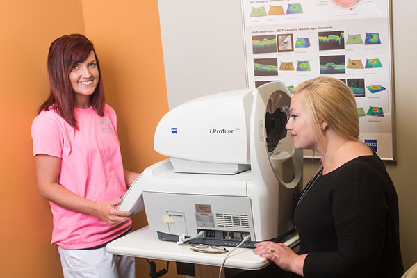
Our OCT helps us better manage glaucoma and diseases of the retina because this technology allows the eye doctor to see the deep tissue layers in the eye. These high-definition images are the only way that they can actually see beneath the surface to the nerve fiber layers where damage occurs. Up until now, eye doctors had to use other tests to indicate damage in this critical area of sight. Common eye diseases such macular degeneration, diabetic retinopathy, and glaucoma are detected early by the OCT when the diseases can be more effectively treated. service.
Visual Fields
Visual Field testing can help save vision because it is another test used to diagnose or rule out glaucoma and other neurological disorders that affect vision. This simple, but effective service has saved lives by detecting various medical conditions such as strokes, brain tumors, and other neurological defects.

i.Scription
i.Scription by Zeiss is a state of the art refraction instrument that uses new wavefront technology to accurately record how light enters your eye to determine a much more precise prescription than ever before.
Optomap Retinal Imaging
A high-definition digital image of the retinal area helps your eye doctor diagnose and manage eye diseases in the delicate retinal area. Damage to these delicate structures of the retinal area is often the first sign of systemic diseases such as MS, diabetes and more. The retina is the “window to the body” and routine retinal imaging can help your eye doctor monitor the changes in your eye health from year to year. New technology from Optos enables the doctor to see a much wider view of the back of the eye compared to traditional retinal imaging.
Corneal Topography
Corneal topography is a computer assisted diagnostic tool that creates a three-dimensional map of the surface curvature of the cornea. The cornea (the front window of the eye) is responsible for about 70 percent of the eye’s focusing power. An eye with normal vision has an evenly rounded cornea, but if the cornea is too flat, too steep, or unevenly curved, less than perfect vision results. The greatest advantage of corneal topography is its ability to detect irregular conditions invisible to most conventional testing.
Corneal topography produces a detailed, visual description of the shape and power of the cornea. This type of analysis provides your doctor with very fine details regarding the condition of the corneal surface. These details are used to diagnose, monitor, and treat various eye conditions. They are also used in fitting contact lenses and for planning surgery, including laser vision correction. For laser vision correction the corneal topography map is used in conjunction with other tests to determine exactly how much corneal tissue will be removed to correct vision and with what ablation pattern.
Cataract Surgery & Low Vision
A cataract is a clouding of the lens inside the eye. The lens sits just behind the pupil and is responsible for focusing light. As the lens increases in cloudiness, it becomes more difficult to see objects clearly and bright lights can cause glare. The most common form of cataract is that of aging and it is inevitable as we mature. In most cases, cataracts do not cause any harm to the eye and cataract surgery is only done to improve the vision in which glasses no longer can improve the vision. Cataract surgery is more the patients decision than ours, because we usually recomend surgery only when the cataracts are affecting your daily life. There is no harm in delaying surgery if your life is only minimally affected.
Our Relationship with local Specialists keeps you seeing your best.
